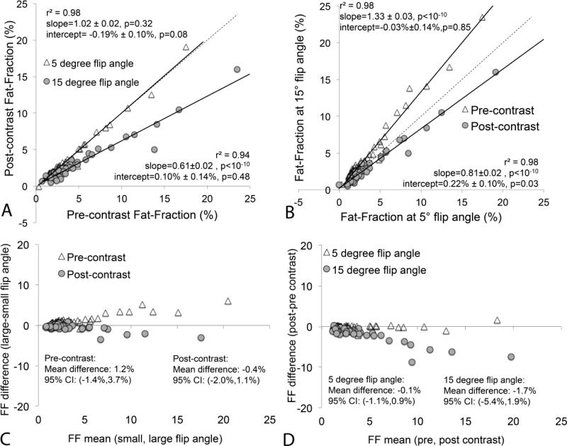
Figure 2.
Scatterplots demonstrate hepatic fat-fractions obtained at low (5°) and high (15°) flip angles before and after the administration of gadoxetic acid. (a) Hepatic fat-fraction is not affected by contrast when T1 related bias is avoided at low flip angles. (b) At high flip angles, hepatic fat-fraction is overestimated prior to contrast and underestimated after contrast administration. This implies that, in the presence of gadoxetic acid in the liver, the T1 of water becomes shorter than the T1 of fat. The dashed line is the line of unity. (c) Bland-Altman analysis comparing low flip angle to high flip angle FF estimates, both before and after contrast. (d) Bland-Altman analysis comparing FF estimates before and after contrast, both using low and high flip angle acquisitions. Note the excellent agreement of T1-independent (ie: low flip angle) fat quantification performed before and after contrast administration.

