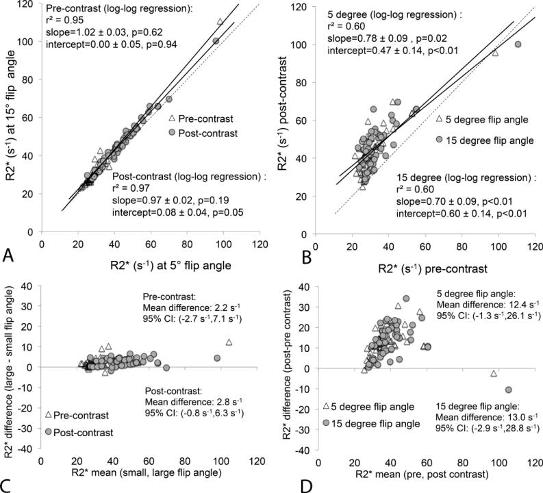
Figure 4.
Comparison between hepatic R2* obtained at low (5°) and high (15°) flip angles before and after the administration of gadoxetic acid. There is no substantial difference in hepatic R2* at low and high flip angles; however, there is significant increase in hepatic R2* following contrast administration at both flip angles. (a) Scatterplot of R2* acquired with low vs high flip angle (both pre- and post-contrast). (b) Scatterplot of R2* pre-contrast vs post-contrast (for low and high flip angles). Dashed line equals the line of unity. (c) Bland-Altman analysis comparing low flip angle to high flip angle R2* estimates, both before and after contrast. (d) Bland-Altman analysis comparing R2* estimates before and after contrast, both using low and high flip angle acquisitions.

