
Figure 1. Abhd5 Suppresses EMT and Growth Advantage of CRC Cells.
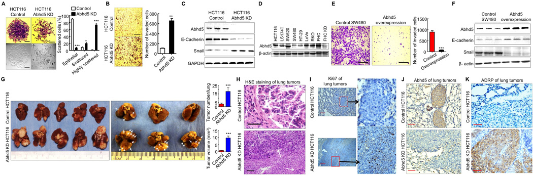
(A) Morphology (crystal violet staining and phase-contrast) of HCT116 control and Abhd5-knockdown (KD) cells. One hundred cells in each colony were counted under microscopy (200x). Ten colonies were counted for each group. Epithelial: cells attached to each other tightly; Scattered: cells scattered, but still attached to others; Highly scattered: the scattered cells that had no contact with others. *P < 0.01. ***P < 0.0001. Scale bar = 50 µm.
(B) Transwell assays. Scale bar = 200 µm.
(C) Western blots of EMT markers.
(D) Western blots of Abhd5 in colon cancer and normal colon epithelial cell lines.
(E) Transwell assays of SW480 cells overexpressing Abhd5. Scale bar = 200 µm.
(F) Western blots of EMT markers in SW480 cells overexpressing Abhd5.
(G) Lung tumor lesions induced by the tail vein injection of HCT116 cells in nude mice (left panel, nonfixed organ; right panel, formalin-fixed organ).
(H) H&E staining of the above lung tumors. Scale bar = 100 µm.
(I–K) Immunohistochemistry of Ki67, Abhd5 and ADRP in the above lung tumors. Scale bar = 100 µm.