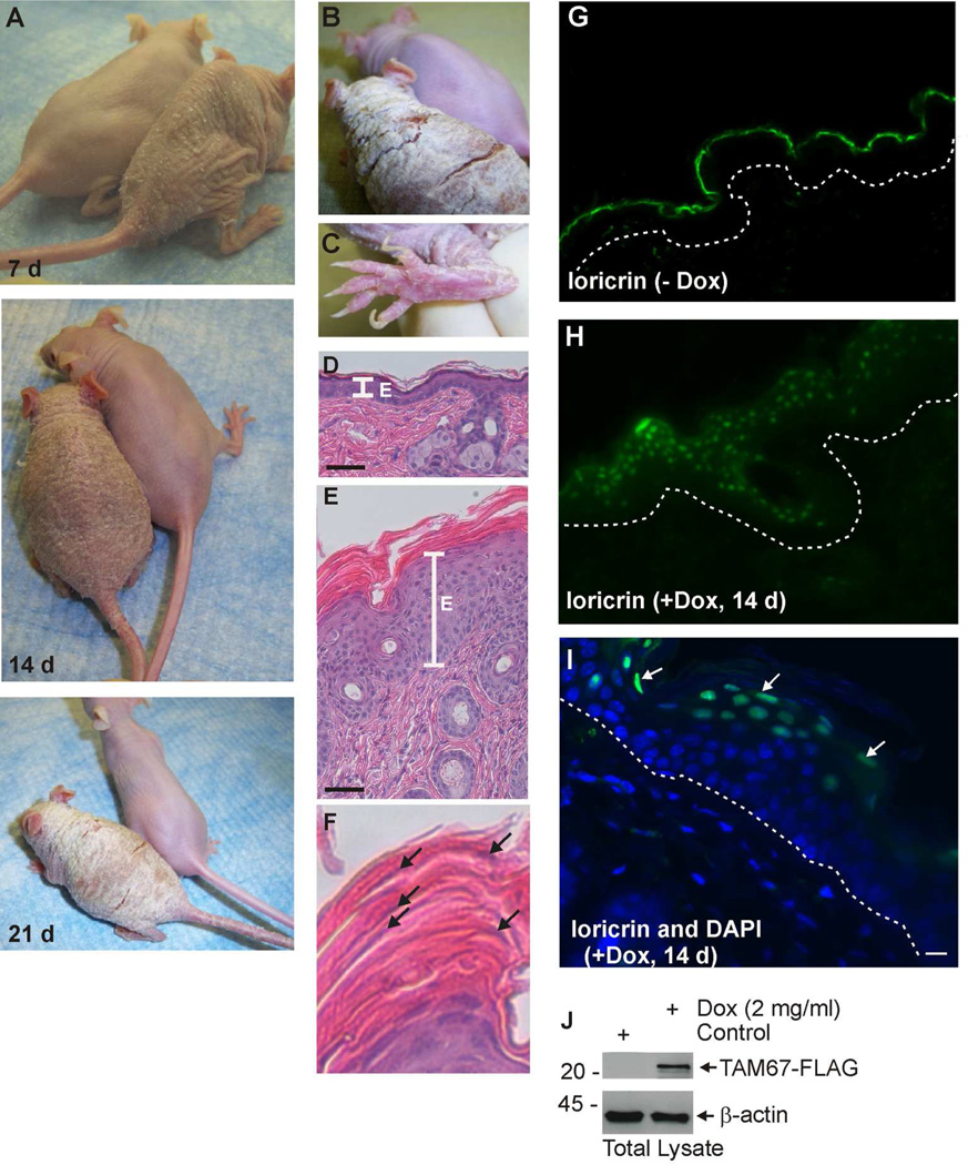
Fig. 2. Characterization of SKH1-TAM67-rTA mice.
SKH1-TAM67-rTA mice were administered drinking water supplemented with or without 2 mg/ml doxycycline. A Doxycycline-treated and untreated littermates were photographed at 7, 14 and 21 d. B/C Enlarged images showing hyperkeratinization of the dorsal epidermis and foot in mice treated for 21 d with doxycycline. Panel B includes an image of an untreated littermate. D Histological appearance of epidermis from untreated littermate. E Histological appearance of epidermis from 14 d doxycycline-treated littermate. The line labeled “E” marks the extent of the living epidermis. F Histological appearance of the cornified layer from panel E shows incomplete nuclear destruction (parakeratosis) in the cornified layer. The arrows indicate nuclei. G/H Loricrin distribution in untreated and 14 d doxycycline-treated SKH1-TAM67-rTA mice. The dotted lines indicate the dermal/epidermal junction. I Co-staining with DAPI (nuclear) and anti-loricrin confirms loricrin nuclear distribution in epidermis from 14 d doxycycline-treated mouse. J Immunoblot shows accumulation of TAM67-FLAG in 14 d doxycycline-treated mice and the absence of expression in untreated littermate.

