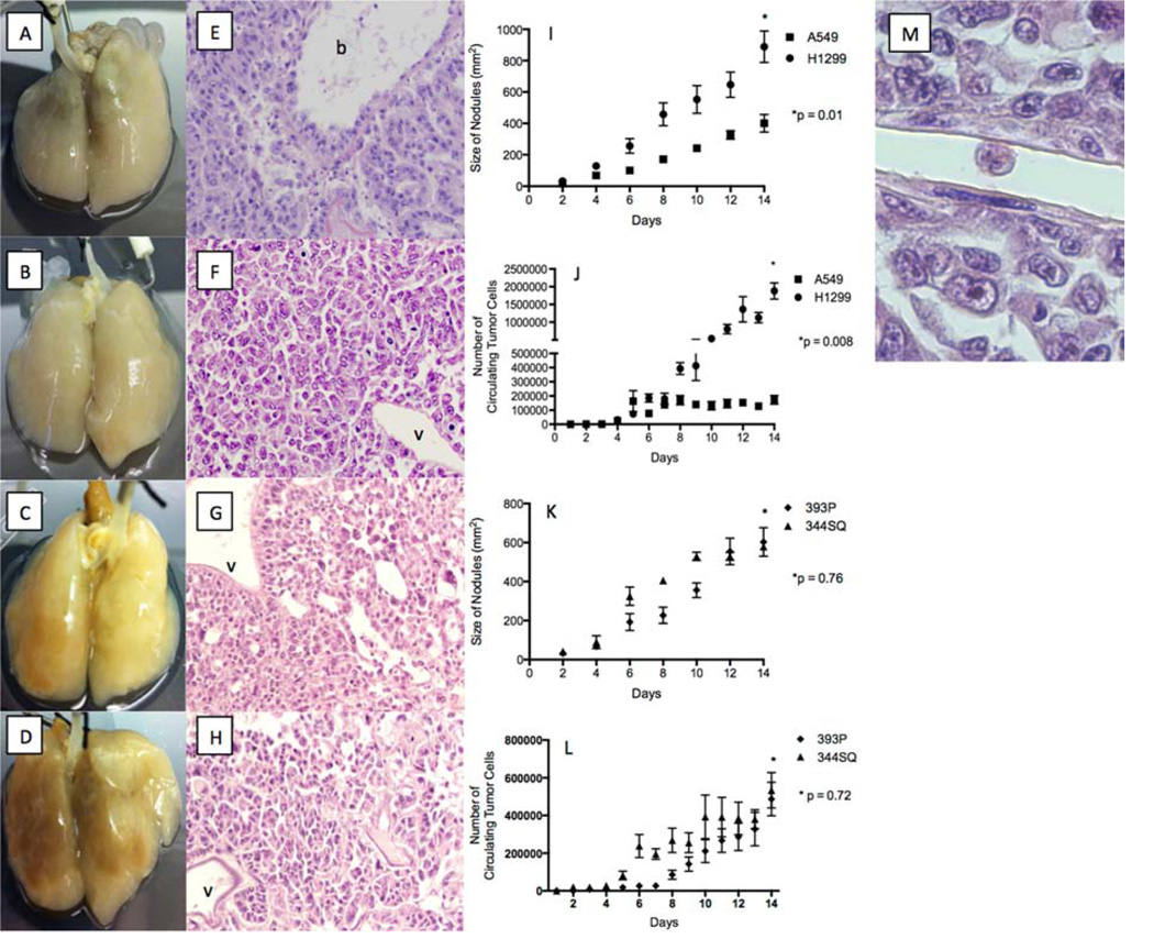
Figure 1. Tumor nodule and CTC formation on 4D model.
(A–D) Image of A549 (A), H1299 (B), 393P (C), and 344SQ (D) cells grown on the 4D model on day 14 showing the presence of tumor nodules. (E–H) Hemotoxyline and eosin staining of the lung tissue from the 4D model on day 14 seeded with A549 (E), H1299 (F), 393P (G), and 344SQ (H) cells show a lack of tumor growth in vasculature (v) and the presence of tumors in the bronchus (b). (I) Bigger tumor nodules on the 4D model seeded with H1299 cells on day 14 compared to the 4D model seeded with A549 cells (p = 0.01). (J) Higher number of live CTCs in the 4D model seeded with H1299 tumor cells as compared to those seeded with A549 tumor cells on day 14 (p < 0.008). (K) No significant difference in tumor nodule size on day 14 between the 4D model seeded with 344SQ and 393P cells (p = 0.76). (L) No significant difference in the number of CTCs on day 14 between the 4D model seeded with 344SQ and 393P cells (p = 0.72). The error bar represents the standard error of mean. (M) Hemotoxyline and eosin staining of lung tissue from the 4D model seeded with H1299 cells, with the live tumor cell in the vasculature. The tumor cell is oval in shape; without any cytoplasmic extension; and with a high nucleus-to-cytoplasm ratio, irregular nuclear contours, and clumpy chromatin.

