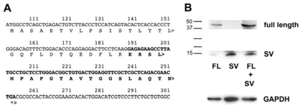
Figure 3.

Expression testing of SV protein. (A) Predicted protein following exon skipping; bold indicates amino acids not encoded by full-length EKLF. (B) Plasmids expressing either FLAG-tagged FL or SV Eklf were transfected as indicated into COS7 cells. Inclusion of MG132 to inhibit proteasome degradation of the small SV product had no effect (not shown). Molecular weight markers are on left; anti-GAPDH was monitored as a loading control (bottom). Exposure of the bottom half of the gel was ~30x greater than the top half.
