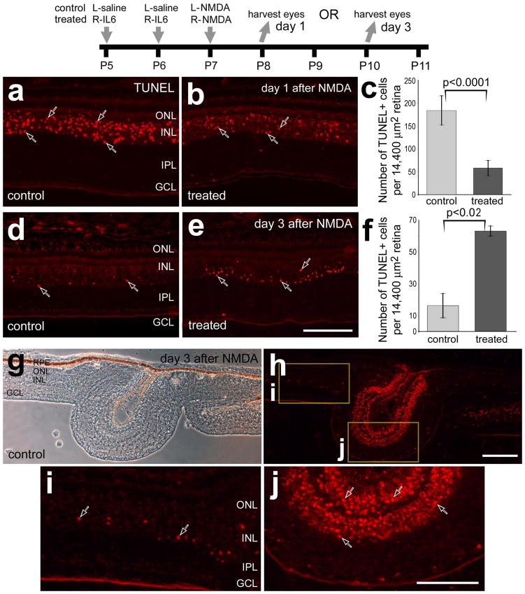
Figure 1.
IL6 delays cell death in NMDA-damaged retinas. Retinas were obtained from eyes that were injected with IL6 (or saline) at P5 and P6, NMDA at P7, and harvested at P8 (1 day after NMDA; a–c) or at P10 (3 days after NMDA; d–j). Vertical sections of the retina were fluorescently labeled in situ for fragmented DNA (TUNEL; a,b,d,e and h–j) or images were taken using phase contrast imaging (g). Hollow arrows indicate TUNEL-positive nuclei. The boxed areas in panel h are enlarged 3.5-fold in panels i and j. Panels c and f are histograms illustrating the mean (±SD) number of TUNEL-positive nuclei per 14,400 μm2 in retinas treated with saline/NMDA or IL6/NMDA. Significance (*p<0.02, **p<0.0001) of difference between control and treated groups was determined using a two-tailed, unpaired t-test. The scale bar (50 μm) in panel e applies to a, b, d and e, the bar in g applies to g and h, and the bar in j applies to j and i. Abbreviations: ONL – outer nuclear layer, INL – inner nuclear layer, IPL – inner plexiform layer, GCL – ganglion cell layer, RPE- retinal pigmented epithelium.

