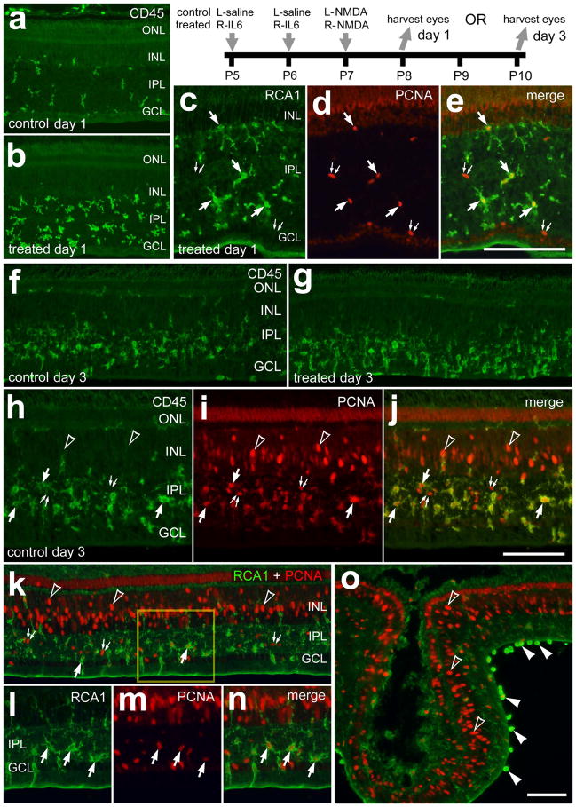
Figure 2.
IL6-treatment accelerates the accumulation of reactive microglia and marcrophage in NMDA-damaged retinas. Retinas were obtained from eyes that were injected with IL6 (or saline) at P5 and P6, NMDA at P7, and harvested at P8 (1 day after NMDA; a–e) or P10 (3 days after NMDA; h–o). Vertical sections of the retina were labeled with RCA1 (green; c, e, k, l, n and o), antibodies to CD45 (green; a, b, f–h and j) or antibodies to PCNA (red; d, e, i–k and m–o). The arrows indicate microglia/macrophage, small double-arrows indicate NIRG cells, hollow arrow-heads indicate the nuclei of Müller glia, and solid arrow-heads indicate presumptive macrophages at the vitread surface of the retina. The scale bar (50 μm) in panel c applies to c–e, the bar j applies to h–j and the bar in o applies to a, b, f, g, k and o. The boxed-out area in k is enlarged 2-fold in l–n. Abbreviations: ONL – outer nuclear layer, INL – inner nuclear layer, IPL – inner plexiform layer, GCL – ganglion cell layer, PCNA – proliferating cell nuclear antigen.

