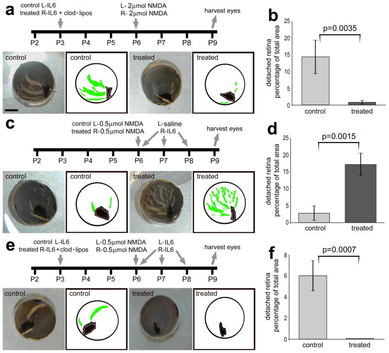
Figure 5.
Activation of microglia/macrophages with IL6 increases retinal detachments and folds, whereas the ablation of microglia/macrophages prevents the formation of retinal detachments and folds. Experimental paradigms for injections of IL6, clodronate-liposomes and NMDA are outlined for each data set (a, c and e). Images include digital photographs and digital tracings of the eyecup circumference (black), pecten (black) and retinal folds/detachments (green; a, c and e). The scale bar (5 mm) in panel a applies to a, c and e. The histograms in panels b, d and f illustrate the mean (± SD) percentage of retinal area that is detached/folded in the different experimental paradigms. The significance of difference (*p<0.005, **p<0.0001) was determined by using a two-tailed, unpaired t-test. Percentage area of retinal folds and detachments was determined from digital micrographs. The detached areas appeared as opacities that were digitally traced and measured by using ImagePro 6.2. The detached retinal area was calculated as a percentage of total retinal area without compensating for concave shape of the eyecup.

