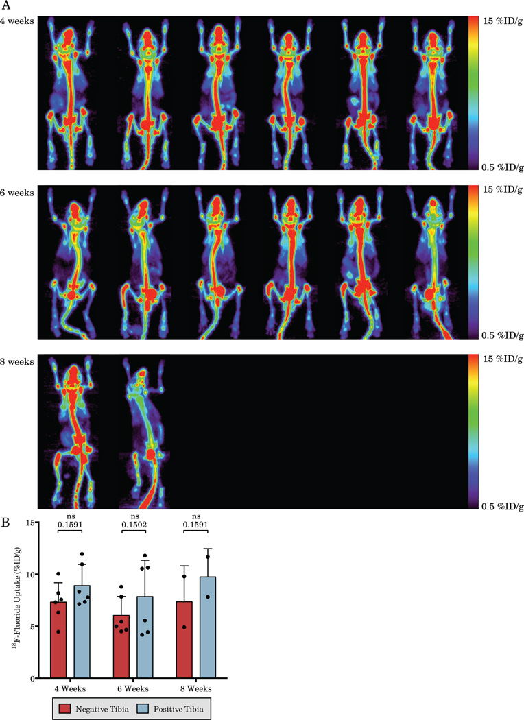
Figure 1.
Serial 18F-Fluoride bone scans of mice bearing LAPC-9 intratibial xenografts (A) and quantification of the positive and negative tibias (B). Clear determination of increased signal in tumor bearing tibias is difficult due to the large degree of non-specific uptake. Each column displays serial imaging of the same mouse and each is matched with the corresponding A11 immunoPET in Figure 2.

