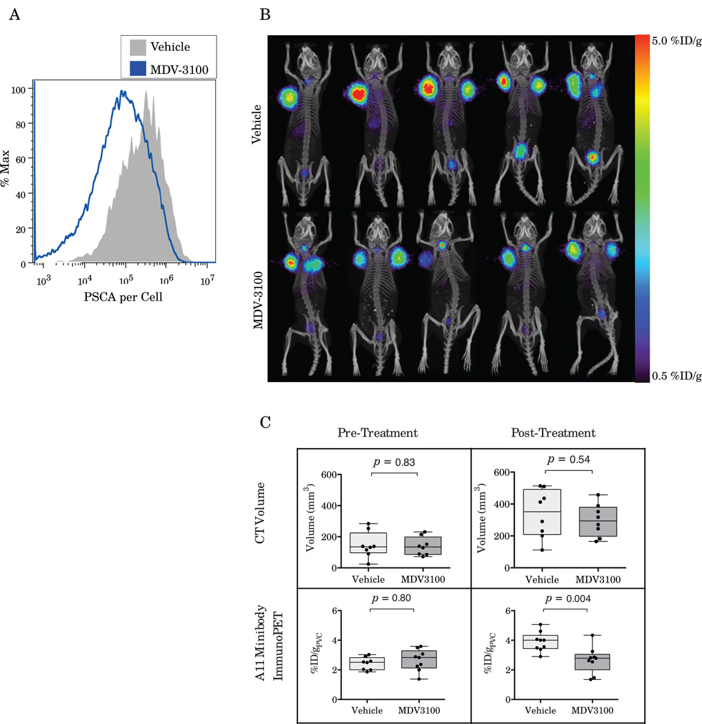
Figure 4.
Ex vivo quantitative flow cytometry shows downregulation of PSCA on LAPC-9 tumor cells in response to treatment with MDV-3100 (A). A11 minibody imaging of mice bearing LAPC-9 xenografts treated with MDV-3100 shows decreased tumor uptake in vivo as compared to vehicle controls (B). Quantification of the microPET/CT results shows no difference in tumor volumes or minibody uptake pre-treatment (C). However, following a week of treatment, A11 immunoPET shows significantly decreased uptake in MDV-3100 treated mice compared to vehicle controls (p=0.004) whereas CT volume shows no difference between the groups (p=0.54).

