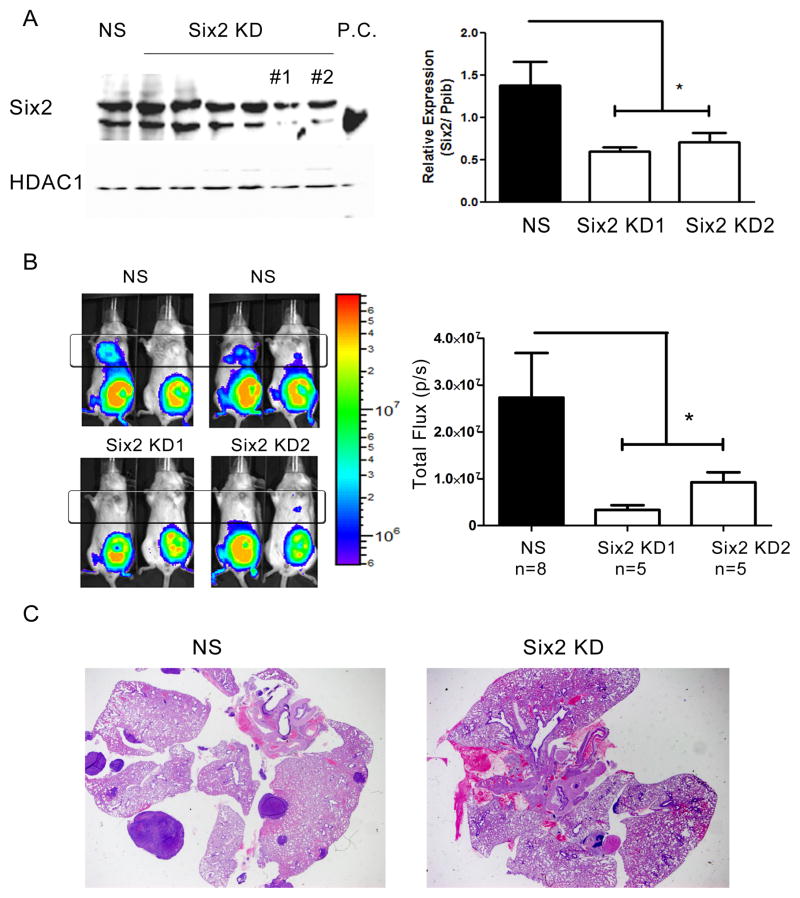
Fig 2. Six2 KD decreases metastasis in the 66cl4 mammary carcinoma models.
(A) 6 different shRNA against Six2 was used to knockdown Six2 in the 66cl4 cells and the most efficient Six2 knockdown cells (KD#1 and KD#2) were used for subsequent studies. Six2 expression was determined using Western blotting (left) and real-time PCR (right) in control (NS, non-silencing) and Six2 KD cells. P.C. stands for positive control, and demonstrates where the Six2 specific band runs. (B) Representative bioluminescent imaging of Balb/c mice injected with control (NS) or Six2 KD cells into the 4th mammary fat pad (left). Quantitation of distant luminescent signal, likely in lungs (boxed region) reveals a significant decrease in metastasis when Six2 is knocked down (right). (C) Histological confirmation of lung metastasis by H& E staining from control and Six2 KD.

