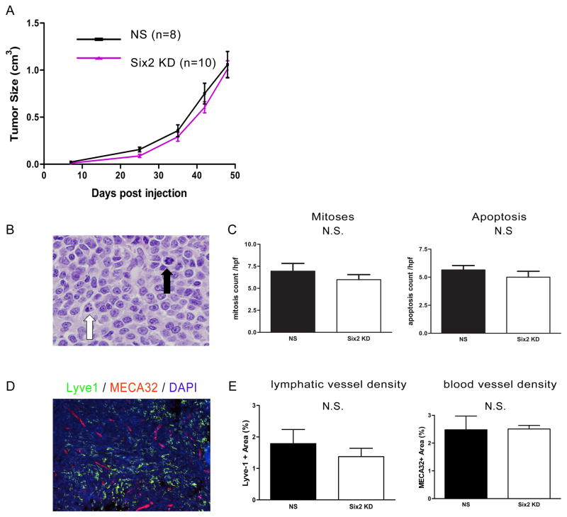
Fig 3. Six2 KD does not affect primary tumor growth.
(A) Primary tumor size in 66cl4 control (NS) injected mice and Six2 KD injected mice (two different shRNA lines were used and combined in the figure) tumors. Tumor size in the animals was measured using calipers, and calculated according to the formula V=1/2(W)(W)(L). (B) Representative histology showing cells undergoing apoptosis or mitosis. Black arrowhead: mitotic cell; white arrowhead: apoptotic cell. (C) Quantification of mitotic and apoptotic cells from 66cl4-NS and 66cl4-Six2 KD tumors. Mitotic and apoptotic cells were counted under ten high power fields per H&E section. Four control and five Six2 KD tumors were counted. (D) Representative picture showing lymphatic vessels using Lyve-1 staining and blood vessels using MECA-32 staining in a 66cl4 tumor. (E) Slidebook software was used to quantify lymphatic or blood vessels in four 66cl4-NS and four 66cl4-Six2 KD tumors. N.S. stands for no statistic significance.

