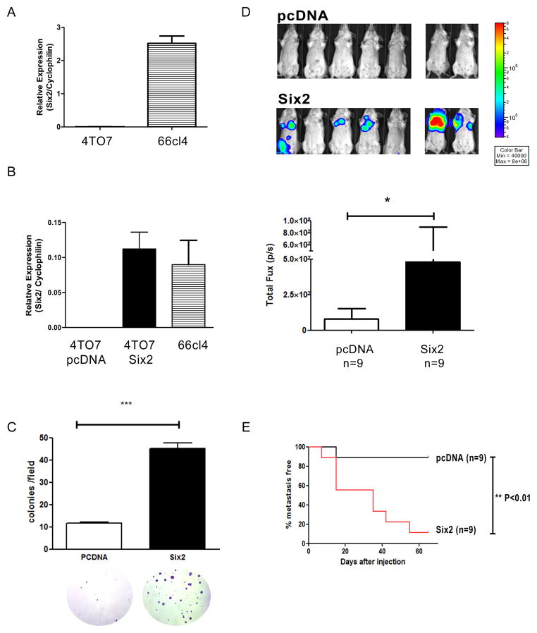
Fig 4. Six2 expression promotes metastasis in the 4TO7 mammary carcinoma model.
(A) Six2 mRNA expression was measured using real-time RT-PCR in 4TO7 and 66cl4 cell lines. (B) pcDNA control or Six2 expressing vectors were transfected into 4TO7 cells and clones were pooled after hygromycin selection. Six2 over-expression in 4TO7 cells was measured by real-time PCR to compare endogenous levels of Six2 in 66cl4 cells to those obtained with ectopic expression of Six2 in 4TO7 cells. (C) Six2 expression increases anchorage independent cell growth. Quantification of colonies formed in soft agar by 4TO7-pcDNA or 4TO7-Six2 cells (upper) and representative pictures of colonies in 4TO7-pcDNA and 4TO7-Six2 cells (bottom). (D) Luciferase labeled 4TO7-pcDNA or Six2 cells were injected into Balb/c mice through the tail vein and in vivo metastasis was measured using IVIS imaging. Representative pictures of animals injected with 4TO7-pcDNA or 4TO7-Six2 cells (top). Quantification of whole body luciferase per animal in 4TO7-pcDNA (n=9) and 4TO7-Six2 (n=9) groups (bottom). (E) Kaplan-Meier plot shows % of metastasis free mice in 4TO7-pcDNA control and 4TO7-Six2 expressing groups. Statistical analysis was performed using the log-rank test.

