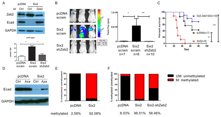
Fig 6. Six2 represses E-cadherin expression via multiple mechanisms.
(A) Zeb2 KD in 4TO7-Six2 cells reverses E-cadherin repression (Top). Expression of Zeb2 and E-cadherin in 4TO7-pcDNA, 4TO7-Six2 and 4TO7-Six2-Zeb2 KD cells was measured by Western blotting. shRNA targeting Zeb2 was delivered into 4TO7-Six2 cells and stable KD was selected using puromycin. Scramble shRNA was delivered into 4TO7-pcDNA and 4TO7-Six2 cells to serve as KD control. Zeb2 KD in 4TO7-Six2 expressing cells decreases anchorage independent cell growth (Lower). Quantification of colony numbers formed by 4TO7-pcDNA, 4TO7-Six2, 4TO7-Six2-Zeb2 KD cells in soft agar. **, P<0.01. (B) Luciferase labeled 4TO7-pcDNA, 4TO7-Six2 or 4TO7-Six2-Zeb2 KD cells were injected into Balb/c mice through the tail vein and in vivo metastasis was measured using IVIS imaging. Representative pictures (left) and quantification (right) of luciferase signal in animals injected with 4TO7-pcDNA (n=8), 4TO7-Six2 (n=8) or 4TO7-Six2-Zeb2 KD (n=7) cells are shown. (C) Kaplan-Meier plot shows overall survival of the injected mice. Statistical analysis was performed using the log-rank test. (D) Restored E-cadherin expression by treating 4TO7-Six2 cells with the DNA methylation inhibitor, 5Aza. 4TO7-pcDNA and Six2 cells were treated with DMSO (vehicle control) or 5Aza (10uM) for 48hrs and whole cell lysates were collected and Western blotting was performed for E-cadherin expression. (E) Six2 expression significantly increases E-cadherin promoter methylation. Genomic DNA from 4TO7-pcDNA and 4TO7-Six2 cells was collected and E-cadherin promoter CpG methylation status was detected using EpiTech Methyl II PCR primer assay (Qiagen) for the mouse Cdh1 promoter. (F) Zeb2 KD in 4TO7-Six2 cells decreases CpG methylation of the Cdh1 promoter. Relative amount of methylated and unmethylated E-cadherin promoter was detected using EpiTech Methyl II PCR primer assay for mouse Cdh1 in 4TO7-pcDNA, Six2, and Six2-Zeb2 KD cells. UM:un-methylated DNA. M: methylated DNA.

