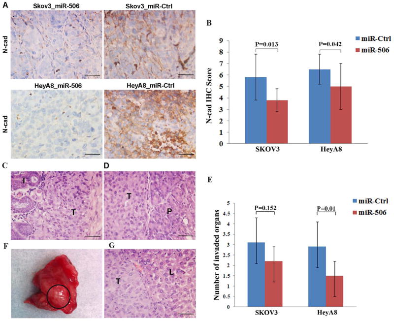
Figure 5. Systemic delivery of miR-506 suppresses invasion and metastasis of OvCa cells in mouse models.
HeyA8-IP2 or SKOV3-IP1 cells were injected into the peritoneal cavities of nude mice. The mice were treated with miRNA incorporated in DOPC nanoliposomes (intraperitoneal administration): control miRNA/DOPC or miR-506/DOPC for each cell line (n = 10 mice per group), for 4–6 weeks. A and B. MiR-506 treatment led to decreased N-cad expression. Representative images of immunohistochemical staining for N-cad in 2 mouse models (A). The statistical analysis shows that N-cad expression is lower in the miR-506 treatment groups than in the miR-Ctrl treatment groups (B). C. SKOV3-IP3 cells invaded the intestines through the mesentery in the mice treated with miR-Ctrl. I, intestine; T, tumor cells. D. HeyA8-IP2 cells invaded the pancreases in the mice treated with miR-Ctrl. P, pancreas; T, tumor cells. E. The statistical analysis shows that the mean number of invaded organs by tumor cells in the miR-506 treatment groups is lower than in the miR-Ctrl treatment groups. F and G. Liver metastasis was found in 2 mice with HeyA8-IP2 cells and treated with miR-Ctrl. Shown is liver metastasis with no peri-hepatic or capsular invasion (F). H & E images of liver metastasis of HeyA8-IP2 cells (G). L, liver; T, tumor cells. Scale bars represent 50 μm.

