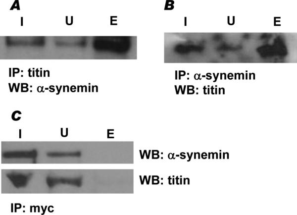
Figure 4. Endogenous α-synemin and titin interact in vivo.

A, Endogenous α-synemin and titin were co-immunoprecipitated from HL-1 cells using anti-titin antibody and visualized using anti-α-synemin antibody (E). B, In reciprocal experiments anti-α-synemin antibody was used to immunoprecipitate endogenous proteins and anti-titin antibody was used to visualize the co-immunoprecipitated titin protein (E). C, In control experiments, α-synemin or titin did not co-immunoprecipitate with anti-myc antibody. In each experiment, the entire volume of the eluate was loaded on the gel along with equivalent volumes of input (I) and unbound (U) fractions: I, input; U, unbound, E, eluate.
