Figure 1.
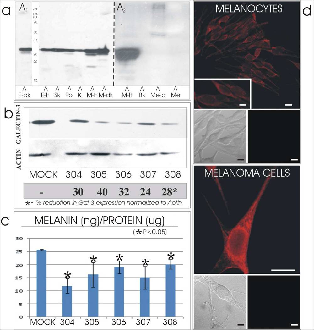
[A] Expression and secretion of galectin-3 by cutaneous cells and tissues. [A1] Galectin-3 of 30 kDa was identified by immunocytochemistry in lysates of dark (E-dk) and light (E-lt) neonatal foreskins and in cultured cells of skMel melanoma (Sk), dermal fibroblast (Fb), keratinocytes (K) and melanocytes derived from light (M-lt) and dark (M-dk) skin. [A2] Media collected from light skin derived melanocytes before (Me) or after (Me-a) acetone precipitation did not exhibit the 30 kDa galectin-3. However the latter did exhibit a 45–50 kDa triplet and a 250 kDa doublet. [Bk = blank lane]
[B & C] Silencing of galectin-3 results in reduced melanin synthesis. Cultured melanocytes derived from one light skin was transfected with control shRNA (Mock) or one of six galectin-3 shRNA (304-8). Amount of [B] galectin-3 protein and [C] melanin content were reduced in thegalectin-3 silenced melanocyte lines by 24–40% and 20–50%, respectively, compared to the Mock transfectants in all melanocyte lines.
[D] Cellular profile of galectin-3. Cultured normal human melanocytes and skMEL melanoma cells demonstrated a prominent perinuclear localization of galectin-3 and minimal dendritic localization. Doublet insets below the immunofluorescent micrographs represent phase (left) and corresponding fluorescent (right) images of cells stained with the galectin-3 primary antibody omitted and secondary used to demonstrate lack of non-specific staining. N=nucleus; C=cytoplasm. Bars = 10 microns; inserts = 5 microns.

