Figure 6.
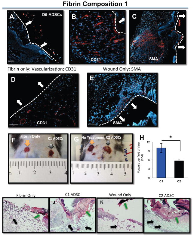
ADSCs remain in the periphery of the wound and within the fibrin gels (A) ADSCs were pre-labeled with DiI and embedded within Composition 1 gels and placed topically over full-thickness mouse wounds; ADSCs remain in the periphery of the wound, and remain encapsulated within the fibrin itself. (B–C) Non-labeled ADSCs were placed topically over full thickness wounds in Composition 1 fibrin gels; CD31 (B) expression and SMA expression (C) is evident when compared to either fibrin only (D) or with the wound only, no treatment condition (E), which are negative for both markers. All secondary antibodies are shown in red. The white bar and white arrows in each of the panels identifies the apex of the healing wound. The white arrows point to the location of the fibrin sealant. (F, G). There are a significantly higher number of vessels per field of view in C1 gels as compared to C2 (H). C1 or C2 gels, respectively, delivered onto full-thickness rodent wounds. In the fibrin-only condition (I), there is not a significant gross histological distinction from the wound-only (K), whereas with C1 gels (J), there is more granulation tissue and cellularity compared to C2 gels (L). Nuclei are counterstained with DAPI (blue). Scale bar = 50 μm

