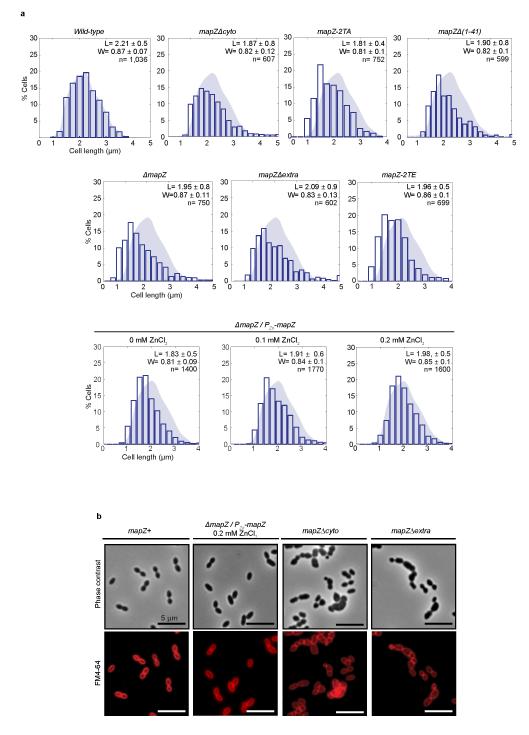
Extended Data Figure 1. Cell-shape analysis of wild-type and mapZ mutants strains.
a. Cell length distribution of wild-type, ΔmapZ, mapZ-2TA, mapZ-2TE, mapZΔcyto, mapZΔextra and mapZΔ(1-41) strains, as well as for ΔmapZ/PZn-mapZ in presence of 0, 0.1 or 0.2 mM of ZnCl2 inducer. Average cell length (L) and width (W) are given with standard deviations for a total of n cells analysed from three independent experiments. For these samples, the two-tailed t distribution P-value determined using non-parametric statistical test was <1.59 10−2, for a critical value of 0.05. b. Phase contrast microscopy and FM4-64 membrane staining imaging of mapZ+ cells (mapZ is restored at the chromosomal locus in ΔmapZ), ΔmapZ/PZn-mapZ (ΔmapZ cells complemented ectopically with PZn-mapZ), mapZΔcyto and mapZΔextra cells. Images are representative of experiments made in triplicate.

