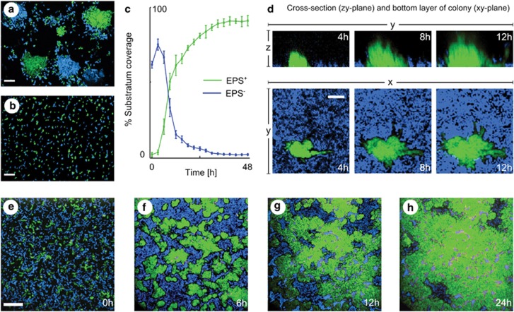
Figure 6.
EPS-producing cells displace non-producing cells from the substratum in a V. cholerae experimental system. (a) A 1:1 mixture of green and blue EPS+ cells form clonal clusters in liquid culture. (b) A 1:1 mixture of blue and green EPS− cells remain dispersed. (c–h) EPS+ (green) in competition with EPS− cells (blue) at different time points. (c) Quantification of surface area coverage by the two strains over time. Bars denote s.e.m., n=3 replicates. (d) EPS+ cells (green) displace V. cholerae EPS− cells (blue) by burrowing under the EPS− strain along the attachment surface in co-culture, even though EPS− cells can remain attached when grown alone (Supplementary Figure S5). (e–h) A time series for the bottom layer of EPS+ (green) competing against EPS− cells (blue). The scale bars in panels a, b and e, f denote 20 μm. The scale bar in panel d denotes 8 μm.

