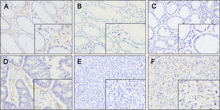
Figure 3. Immunostaining of SPARC in normal and malignant gastric tissues.
(A) Normal gastric tissue showing SPARC expression. The staining was present in stromal fibroblasts and normal epithelium. (B and C) Immunostaining of SPARC was weak in well-differentiated gastric cancer; however, stromal fibroblasts within and surrounding the tumor showed staining of variable intensity. (D) Immunostaining of SPARC was faint or absent in moderately differentiated gastric cancer. (E and F) Immunostaining of SPARC was absent in poorly differentiated gastric cancer and faint in stromal fibroblasts within the tumor. Positive staining of SPARC is indicated by a brown color. ×160 magnification.

