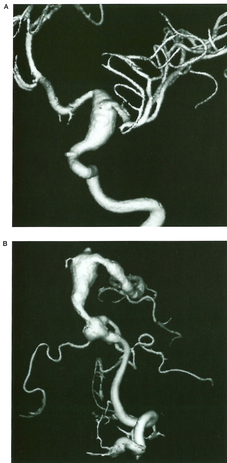
Figure 1.
Case 1, 7-year-old boy with CMCC. A) 3D Angiography of the left internal carotid artery showing a fusiform arterial dilatation of the supraclinoidal internal carotid artery. B) 3D Angiography of the left vertebral artery: large fusiform aneurysm of the basilar artery involving the proximal part of the left posterior cerebral artery, associated with another aneurysm of the same type in the distal part of the left vertebral artery at the vertebrobasilar junction.

