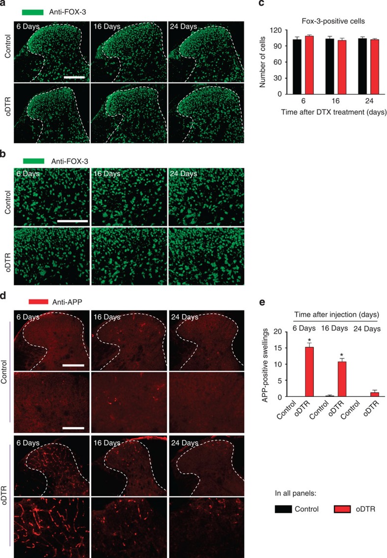
Figure 9. Analysis of neuron-specific markers in mice with inducible oligodendrocyte ablation (oDTR) as compared with control mice.
(a–c) Immunohistochemistry with an antibody-recognizing FOX-3, a neuronal marker, indicates lack of change in neuronal density in DTX-treated oDTR mice as compared with corresponding control mice. spinal dorsal horn and surrounding white matter are shown in a and images in b represent high-magnification images from the grey matter region. The corresponding quantitative analysis of FOX-3-positive cells over an area of 22,500 μm2 is shown in c. (d,e) DTX-treated oDTR mice show an early increase in APP expression, a marker of acute axonal injury via immunohistochemistry; higher-magnification view shown in lower panels. Corresponding quantification of APP immunoreactive axonal swellings over an area of 22,500 μm2 is represented in e. Data were analysed via analysis of variance of random measures followed by post-hoc Fisher’s test; n=3 or 4 mice per group; *P<0.05. Scale bar, 300 μm in a, and d (upper row, low magnification), and 150 μm in b, and d (lower row, high magnification).

