
Figure 1.
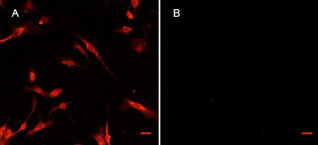
Images under a confocal microscope showing immunohistochemical staining of the intermediate filament and glial fibrillary acidic protein (GFAP) in isolated enteric glia.
(A) and (B) are images of detected fluorescence at 560–610 nm with a laser excitation at 594 nm. The secondary antibody in all cases was to rabbit IgG and was incubated with cells at 1:2 000 for 1 hour, in the dark. The primary antibody was: (A) rabbit anti-GFAP, 1:500; (B) rabbit anti-myelin protein zero, 1:100.
Staining (red, labeled GFAP) in (A) is mostly apparent on cytoskeletal elements surrounding the nucleus and throughout the cytoplasm. While microscope settings are maintained during imaging of (B), no fluorescence is visible excluding a slight signal in the lower left quadrant of (B). Scale bars: 30 μm for both A and B