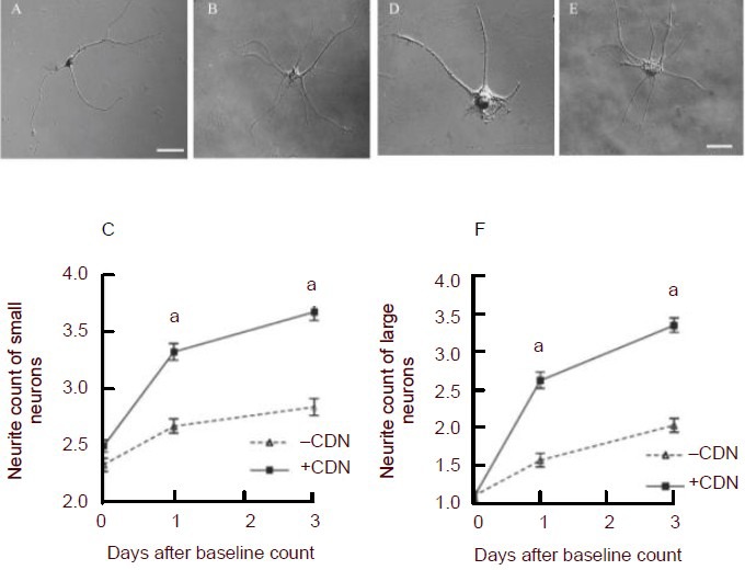
Figure 2.

Examples of small (A, B, C) and large dorsal root ganglion (DRG) neurons (D, E, F) demonstrating the effects of incubation with enteric glia-conditioned medium.
(A) and (B) are differential interference contrast (DIC) images of single, dissociated small DRG neurons, and (D) and (E) are images of single dissociated large DRG neurons under a Nikon Eclipse 80i microscope.
(A) and (D) show typical neurons that had been cultured in neural basal medium. (B) and (E) show neurons that were cultured in enteric glia-conditioned medium. (C) and (F) show average neurite counts (± SEM) in DRG neurons (C, small; F, large) plotted against time.
Squares (+CDN) represent counts taken from DRG neurons incubated in enteric glia-conditioned medium, whereas diamonds (–CDN) represent counts taken from DRG neurons incubated in standard, unconditioned neurobasal medium. aP < 0.001, vs. –CDN. Neurite counts were analyzed using the Mann-Whitney U test with the Bonferroni correction applied. Scale bars: 30 μm for all of the images.
