
Figure 2.
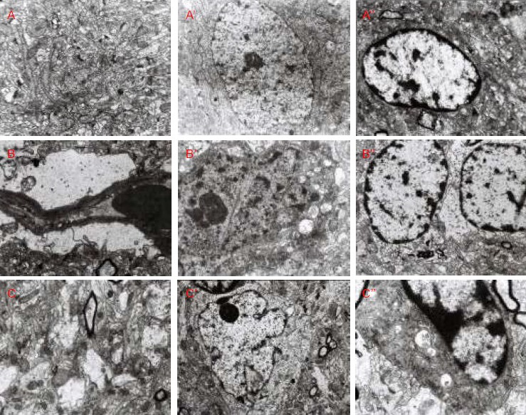
Ultrastructure of rat right parietal cortex (transmission electron microscope).
In the normal control group, rat right cerebral parietal cortical neuropil (A, × 10 000), neurons (A’, × 6 000), and glial cells (A’’, × 7 500) showed normal ultrastructure.
In the low-dose dibutyltin dilaurate group, the capillary ultrastructure was normal (B, × 10 000), but neuronal damage was visible (B’, × 12 000), and glial cells demonstrated cavitation (B’’, × 7 500).
In the high-dose dibutyltin dilaurate group, neuropil cavitation (C, × 10 000), neuronal deformation (C’, × 5 000), and glial cell cavitation (C’’, × 15 000) were visible.