Figure 1.

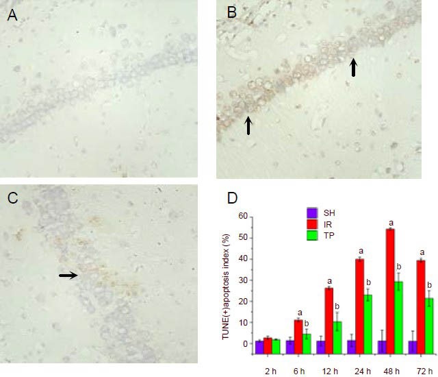
Tea polyphenols reduce apoptosis in the hippocampus induced by cerebral ischemia/reperfusion injury (TUNEL staining, × 400).
Rats were sham treated (SH group), or underwent ischemia/reperfusion (IR group), or underwent ischemia/reperfusion followed by immediate injection of 200 mg/kg tea polyphenols (TP group). Rats were sacrificed at 2, 6, 12, 24, 48 or 72 hours after ischemia/reperfusion, and the apoptotic levels were examined by TUNEL staining. Shown are representative TUNEL staining figures from the hippocampal CA1 region at 48 hours for the SH group (A), IR group (B) and TP group (C).
D shows the percentage of TUNEL-positive apoptotic cells from the three groups at different time points. The hippocampal CA1 region was observed under a light microscope (×400). Five fields of vision were randomly selected to calculate the apoptosis index, defined as the number of apoptotic cells/the total number of cells × 100%.
Arrows indicate TUNEL-positive cells. Under ischemia/reperfusion injury, the apoptotic levels increased significantly at 6 hours and peaked at 48 hours (aP < 0.01, vs. SH group). From 6–72 hours, tea polyphenols administration reduced apoptosis induced by ischemia/reperfusion injury (bP < 0.01, vs. IR group).
Data are presented as mean ± SD, n = 6 per group per time point. Different groups were compared using analysis of variance for two-factor factorial design and least-significant difference t-test.
