Figure 2.

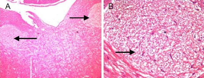
Hematoxylin-eosin stain section of rat brain. Arrow marks point out the damage in the focuses of nucleus of solitary tract and nerve conduction bunch, which were newborn after damage appeared.
(A) × 40; (B) × 100.

Hematoxylin-eosin stain section of rat brain. Arrow marks point out the damage in the focuses of nucleus of solitary tract and nerve conduction bunch, which were newborn after damage appeared.
(A) × 40; (B) × 100.