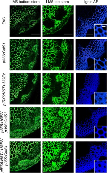
Figure 6.

Galactan and lignin detection in stem sections. β-1,4-galactan was detected by immunofluorescence microscopy using the LM5 antibody. Stem sections from the top and bottom of inflorescence stems were analyzed. Plants co-expressing either pIRX5:NST1-UGE2/ p35S:GalS1 or p35S:UGE2/p35S:GalS1 show a very strong labeling of galactan in the secondary wall as compared to the empty vector control or plants expressing only p35S:GalS1 or pIRX5:NST1-UGE2. Visualization of lignin autofluorescence using a confocal microscope under UV light shows the increase in fiber cell wall density with constructs using the pIRX5 promoter. Bars are 100 μm for bottom stem and lignin autofluorescence pictures, 50 μm for top stems and 10 μm for lignin close ups.
