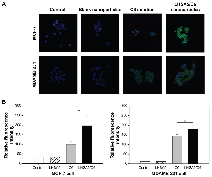
Figure 8.
In vitro cellular uptake of coumarin 6.
Notes: The cellular uptake was observed by (A) CLSM and (B) FACS in MCF-7 and MDAMB 231 cells after incubating for 2 hours. Merged images composed of coumarin 6 (green color) and DAPI (blue color) are shown. Groups were as follows: control, blank LHSA5 nanoparticles, coumarin 6 solution, coumarin 6-loaded LHSA5 nanoparticles. *P<0.05, between two groups.
Abbreviations: FACS, fluorescence-activated cell sorter; CLSM, confocal laser scanning microscope; LHSA, LMWH-SA; LMWH, low-molecular-weight heparin; SA, stearylamine.

