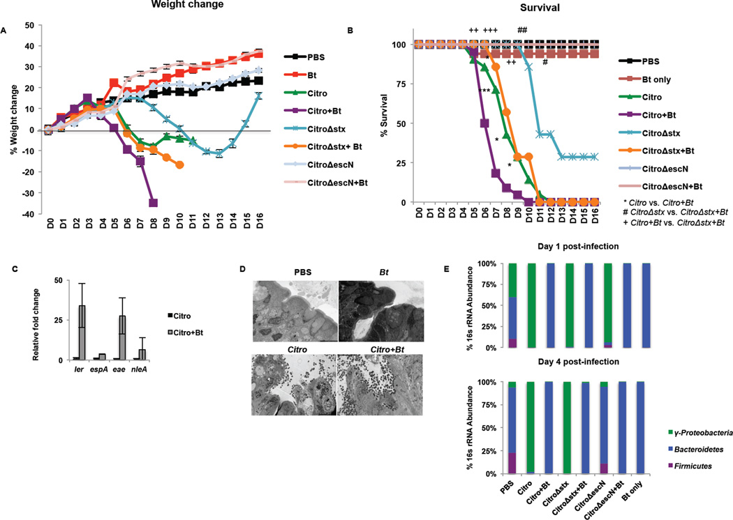
Figure 5. Bt mediates its pro-virulence effect on C. rodentium by enhancing expression of the T3SS, not by a bloom in the C. rodentium population.
C3H/HeJ mice were treated for 5 days with an antibiotics regimen to deplete gut microbiota. Half of the mice were reconstituted with Bt (+Bt) while the remainder of the mice were left deplete of gut microbiota. Mice were mock-infected (PBS, Bt only) or infected with C. rodentium (Citro, Stx+), C. rodentiumΔstx (CitroΔstx, Stx-), or C. rodentiumΔescN (CitroΔescN). A, Weight loss or gain from baseline (weight at day 0) over the course of infection (blue: mock-infected, reconstituted with Bt; red: Citro-infected, deplete of microbiota; green: Citro-infected, reconstituted with Bt; purple: CitroΔstx-infected, deplete of microbiota; turquoise: CitroΔstx-infected, reconstituted with Bt; orange: CitroΔescN-infected, deplete of microbiota; light blue: CitroΔescN-infected, reconstituted with Bt). B, Survival after infection (n = 7–22 mice/group; error bars, s.d.; ***P < 0.001, **P < 0.01: comparison of Citro vs. Citro+Bt; ##P < 0.01, #P < 0.05: comparison of CitroΔstx vs. CitroΔstx+Bt;+++P < 0.001, ++P < 0.01: comparison of Citro+Bt vs. CitroΔstx+Bt). C, qRT-PCR analysis of ler, espA, eae, and nleA from mRNA isolated from fecal pellets of infected animals. Significance is indicated as follows: one asterisk P≤0.05, two asterisks P≤0.01; three asterisks P≤0.001. D, Ultrastructure of the distal colon harvested five days post-infection from mock-infected (PBS, Bt) or C. rodentium-infected mice either deplete of gut microbiota or reconstituted with Bt, 2500×. Microvilli destruction and C. rodentium forming attaching and effacing (AE) lesions on the colonic epithelium can be observed. Original magnification, 2500X (TEM). E, qRT-PCR of 16s rRNA from the major phylogenetic groups (green: Proteobacteria, blue: Bacteroidetes, purple: Firmicutes) from feces collected on day 1 and day 4 post-infection.

