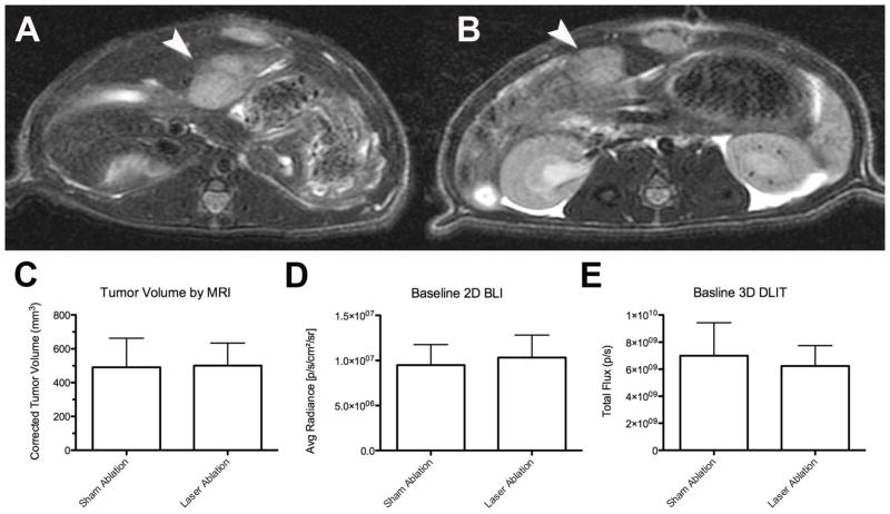
Figure 5.
Baseline N1S1 tumor volume and bioluminescence in sham versus laser ablation groups. N1S1luc2 tumor-bearing rats randomized to sham ablation (N=6) or laser ablation (N=6) underwent pre-ablation non-contrast enhanced 3 Tesla magnetic resonance imaging (MRI; GE Healthcare) to confirm tumor size and location as well as two-dimensional bioluminescence imaging (BLI) and three-dimensional diffuse luminescence tomography (DLIT) using an IVIS200 optical imaging system (Caliper, a PerkinElmer Company) to assess tumor function. Representative pre-ablation axial fast spin echo (FSE) T2-weighted MR images of N1S1 tumors in (A) sham and (B) laser ablation groups demonstrate hyperintense T2-weighted N1S1 tumors (denoted by white arrowhead). (C) Tumor volumes were calculated from MR images, (D) average radiance (photons/sec/cm2/sr) from 2D BLI images and (E) total flux (photons/sec) from 3D DLIT images and compared between sham and laser ablation groups using an unpaired t-test (or Exact Mann Whitney test). There were no significant differences in any of the baseline parameters between groups (p>0.05). Data are presented as mean±SEM.

