Fig. 4.
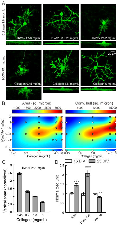
Effects of collagen and IKVAV-PA concentration on PC dendrite morphology. (A) Representative images of PCs (calbindin+), cultured for 16 days on hybrid matrix with different concentrations of collagen and PA. For each condition, the upper image shows top projection, and the lower image shows side projection from a confocal image stack. (B) Colormap representation of the area and convex hull of PC surface dendrites on hybrid matrices with a range of collagen and PA concentration. White circles indicate the sampling points (each point represents an average of 18–71 PCs); at compositions marked by gray circles the matrix became too soft to sustain the staining and imaging procedure. (C) Vertical span of PCs are plotted against the collagen concentration in hybrid matrix (n ≥28 PCs for each concentration). (D) Temporal change in PC dendrite morphology on hybrid matrix (collagen 1.8 mg/mL, IKVAV-PA 1 mg/mL) after16 and 23 days of culture (**:p<0.01, ***:p<0.001, n>50 PCs).

