Fig. 6.

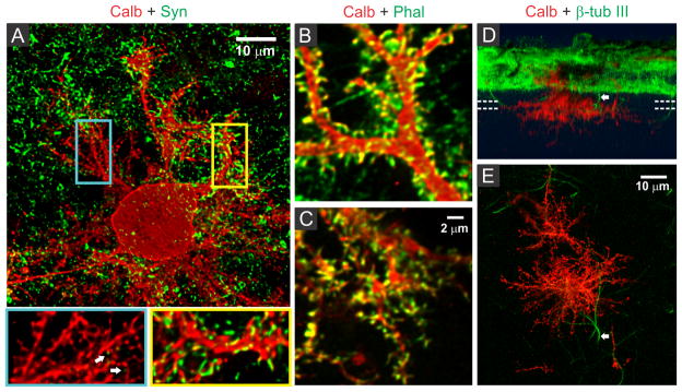
Spine development and synaptic connectivity on PC dendrites. (A) Distribution of presynaptic terminals (synaptophysin, green) on PC dendrites (calbindin, red) in two weeks old culture on hybrid matrix (collagen 1.8 mg/mL, PA 1mg/mL). Magnified views of segments from substrate-penetrating (blue rectangle) and surface dendrites (yellow rectangle) show the presence of numerous spiny protrusions, although the substrate-penetrating dendrites are only rarely associated with presynaptic terminals (white arrow). The spiny protrusions, however, are rich in actin (phalloidin staining) in both surface dendrites (B) and substrate-penetrating dendrites (C). (D) SFP (Simulated fluorescent process) rendered side-view of a confocal image stack showing occasional penetration of non-PC neurites (β-tubulin III+/calbindin-) inside the matrix. (E) Top projection view from a 6.5 μm thick slice volume (indicated by parallel dotted lines in D) captures one such non-PC neurite (arrow) in close proximity to the substrate-penetrating PC dendrites.
