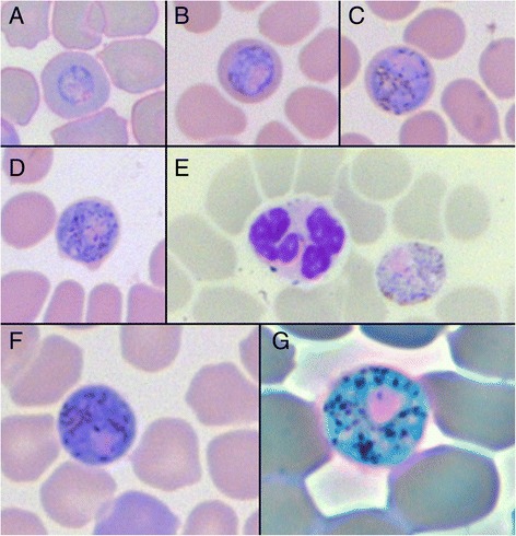
Figure 1.

Images of Polychromophilus murinus gametocytes in Myotis daubentonii erythrocytes. (A-D) Different maturation stages of a gametocyte; (E) Mature male, microgametocyte with leucocyte; (F) Mature female, macrogametocyte; (G) Mature female, macrogametocyte, phase-contrast filter. Images A-F 600x magnification, image G 1000x magnification.
