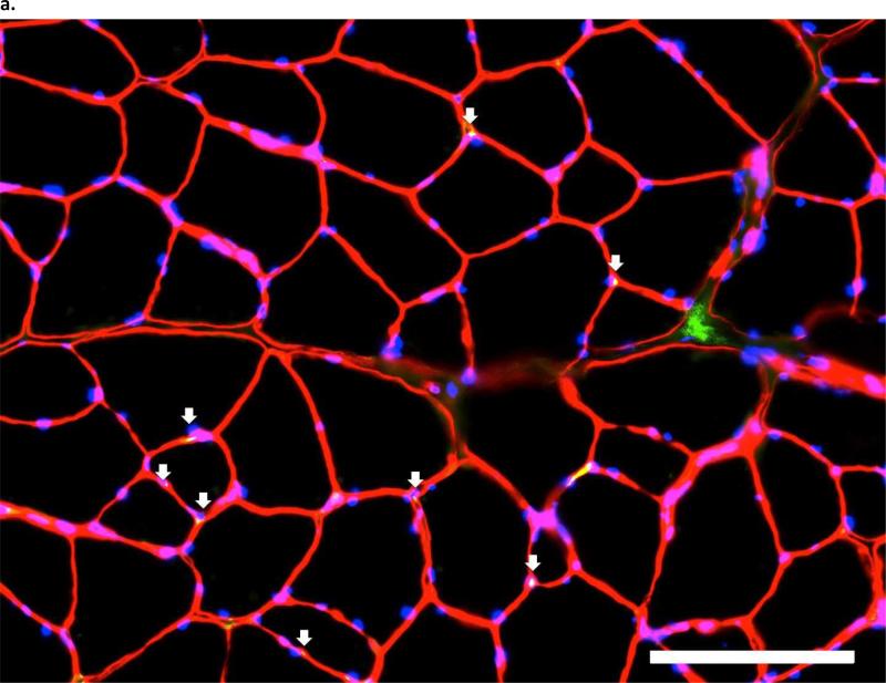
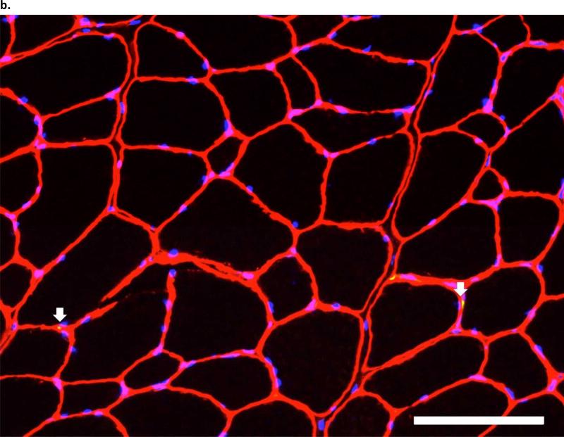
Figure 4. pbhImmunofluorescent images of Pax7 in the third parasternal intercostals, from (a.) an ITTO animal and (b.).
a SHAM animal. Arrows show Pax7-positive nuclei, which were defined as areas that fluoresced with Pax7-AlexaFluor-488, were contained within the sarcolemma and basal membrane, and co-localized with DAPI staining. Each image layer was acquired with exposure times of 150-220 msec and gain of 2.0. At least 250 fibers were analyzed with 10X eyepiece and 20X magnification. Scale bar = 100 μm.


