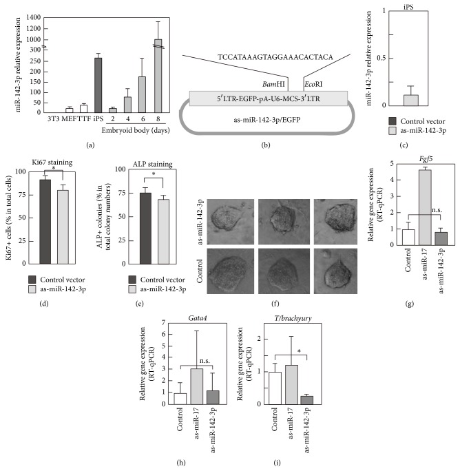
Figure 1.
Differential expression level of miR-142-3p in fibroblasts and iPS. (a) Expression of miR-142-3p was examined by RT-qPCR in various cells. Total RNA was extracted from indicated cells, and RT-qPCR was done using TaqMan MicroRNA systems. U6 shRNA was used as a control. Experiments were done three times using independently prepared cells, and average values with standard deviation are shown. (b) Schematic representation of antisense- (as-) miR-142-3p and EGFP expression plasmid. LTR was used to drive EGFP, and U6 promoter was used to drive as-miR-142-3p. (c) Effect of overexpressed as-miR-142-3p for expression level of endogenous miR-142-3p in iPS cells. as-miR-142-3p/EGFP or control vector was transfected into iPS, and, after 24 hours, level of miR-142-3p in iPS was examined by RT-qPCR. Data were expressed as relative expression level of miR-142-3p in as-miR-142-3p/EGFP expressing cells to that in control vector expressing cells. Experiments were performed three times, and average values with standard deviation are shown. (d, e, and f) Effects of expression of as-miR-142-3p for proliferation and alkaline phosphatase (ALP) expression of iPS. as-miR-142-3p/EGFP plasmid was transfected into undifferentiated iPS, and EGFP positive cells were purified by a cell sorter. Then EGFP positive cells were cultured for 2 days for Ki67 immunostaining and for 5 days for ALP assay. Immunostaining with anti-Ki67 antibody or ALP staining was done, and positive cells were counted under a microscope. Experiments were performed three times, and average values with standard deviation are shown. In (f), morphology of representative colonies of as-miR-142-3p or control vector transfected iPS is shown. (g–i) Expression of lineage marker genes in embryoid body (EB). iPS cells were transfected with as-miR-142-3p/EGFP or as-miR-17/EGFP as a control, purified according to their expression of EGFP, and then subjected to an EB formation. After 6 days of culturing in EB formation condition, the differentiation of cells into the ectodermal (g), endodermal (h), and mesodermal (i) lineages was assessed using RT-qPCR with primers against Fgf5, Gata4, and T brachyury, respectively. P value, ∗ < 0.05 and n.s. > 0.05, was calculated by Student's t-test.

