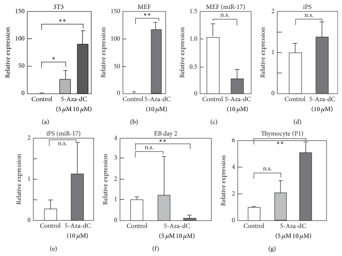
Figure 2.
5-Aza-2′-deoxycytidine (5-aza-dC) treatment upregulates miR-142-3p in fibroblasts. (a–g) 3T3 (a), MEF (b, c), iPS (d, e), embryoid body (EB) formed from mouse iPS (f), or mouse thymocytes (g) were treated with 5-aza-dC at indicated final concentration (5 or 10 μM). Cells were cultured for 3 days in the presence of 5-aza-dC, except for EB, which was treated with 5-aza-dC for two days. Control cells were treated with DMSO. Then, cells were harvested, and total RNA was extracted. Level of miR-142-3p or miR-17 was examined by RT-qPCR. Value of U6 was used as a control. Values are expressed as relative to those of control samples of each cell type and are average of 3 or 4 times experiments with standard deviation. P value, ∗∗ < 0.01, 0.01 < ∗ < 0.05, and n.s. > 0.05, was calculated by Student's t-test.

