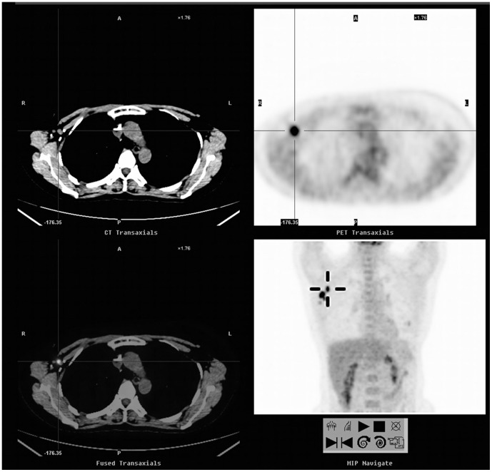
Figure 2. A 46-year-old woman with a history of breast cancer (invasive ductal carcinoma of the right breast, stage IIIA) underwent a FDG-PET/CT scan for asymptomatic follow-up.
FDG-PET/CT showed glucose hypermetabolism in the right axillary nodes (SUVmax = 7.7, cross cursor). The pathological analysis confirmed the presence of metastatic lymph nodes. The patient's serum CA 15-3 remained within normal ranges throughout the course. The patient remained on clinical follow-up at the end of the study.

