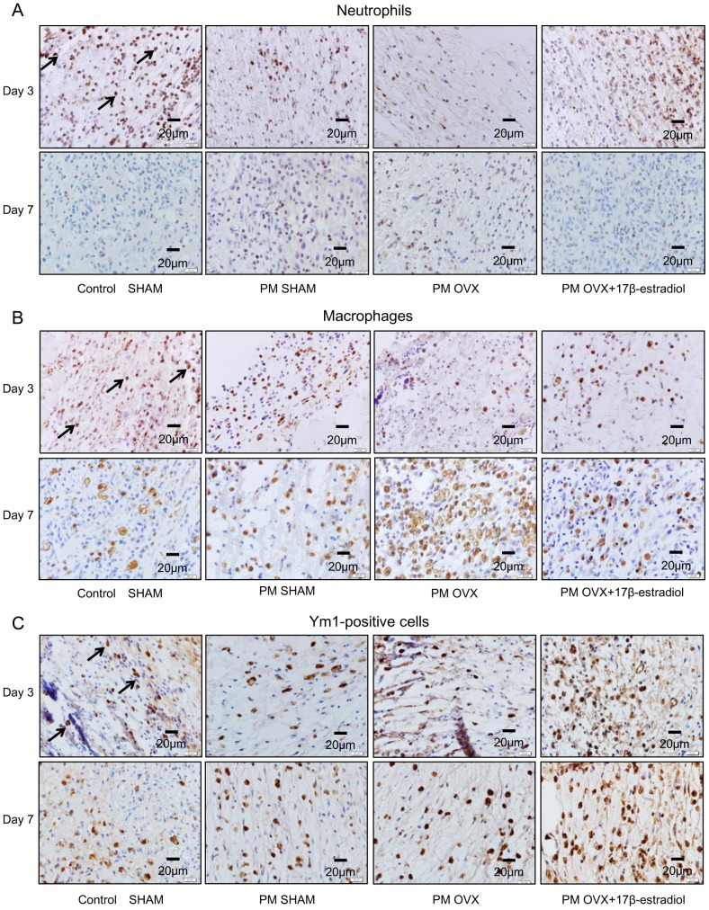
Figure 5. Immunohistological staining for neutrophils, macrophages and Ym1-positive cells on days 3 and 7.
(A) Neutrophils (arrows) stained with anti-neutrophil antibody, (B) macrophages (arrows) stained with anti-Mac-3 antibody and (C) Ym1-positive cells (arrows) stained with anti-Ym1 antibody were observed in wound tissue on days 3 and 7.

