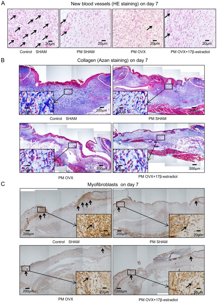
Figure 7. HE staining for new blood vessels, Azan staining for collagen fibers and immunohistological staining for myofibroblasts on day 7.
(A) HE staining on day 7. Note that arrows indicate new blood vessels that show circular structures or include red blood cells. (B) Azan staining on day 7. Collagens stained blue were observed in the granulation tissue. Insets highlight regions of granulation tissue at higher magnification. (C) Myofibroblasts (arrows) stained with anti-α-SMA antibody on day 7. The myofibroblasts appeared at the wound edges on granulation tissue. Insets highlight regions of the wound edge on granulation tissue at higher magnification.

