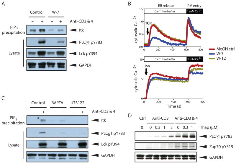
Fig 5. CaM promotes Itk activity and amplifies Ca2+ signaling in a positive feedback loop.
(A) Effect of CaM inhibition (W-7) on TCR-stimulated Itk binding to PI(3,4,5)P3 and Itk-mediated PLCγ1 phosphorylation. Thymocytes were stimulated with antibodies recognizing the CD3 and CD4 (anti-CD3 & 4) subunits of the TCR for 1 minute. TCR stimulation was performed in the presence of vehicle (MeOH ctrl) or W-7 (30μM). Itk association with PI(3,4,5)P3-coated beads and TCR-induced Lck and PLC γ1 phosphorylation at the indicated residues was assessed by Western blot analysis. (B) TCR-induced or ionomycin-induced cytosolic calcium accumulation in thymocytes exposed to vehicle (MeOH ctrl), W-7, or its inactive analog W-12. Ca2+ release from the ER was measured first in the absence of extracellular Ca2+ followed by Ca2+ entry through the plasma membrane (PM) by addition of 1 mM Ca2+ to the sample buffer. TCR stimulation was performed as in (A) in the presence or absence of W-7 (30μM) or W-12 (30μM). (C) Effect of depletion of Ca2+ from the ER by BAPTA-AM (10μM) and EGTA (5 mM) or inhibition of PLCγ1 catalytic activity (U73122, 5μM) on Itk binding to PI(3,4,5)P3 and subsequent Itk-mediated PLCγ1 phosphorylation. Thymocytes were pretreated with indicated reagents for 30 minutes prior to stimulation with biotin-conjugated antibodies to CD3 and CD4 and streptavidin in warm PBS. Samples were separated by SDS-PAGE and analyzed by Western blot analysis with the indicated antibodies. (D) Effect of Thapsigargin (Thap)-mediated increase of cytosolic Ca2+ on Itk-dependent PLCγ1 phosphorylation. Thymocytes were stimulated for 1 minute with biotin-conjugated antibody to CD3 and where indicated CD4 in the presence of 0 – 1 μM Thap. Samples were separated by SDS-PAGE and analyzed by Western blot analysis with the indicated antibodies. All data are representative of 3 experiments.

