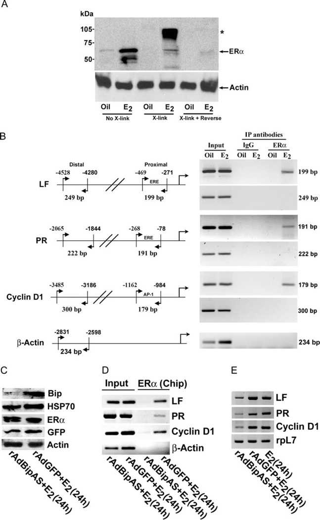
Fig. 4. Analysis of Estrogen-Dependent Effects after Perturbation of Bip Expression in the Uterus.
A, Analysis of estrogen-induced ERα fixation to high molecular complex by Western blotting. Uterine tissues were subjected to cross-linking using 1% formaldehyde (X-link), followed by reversal of cross-linking using heat treatment (X-link + reverse). Tissue extracts at different steps of this procedure were analyzed by Western blotting using ERα-specific antibody. Arrow indicates the position of uncomplexed ERα band, whereas the asterisk (*) denotes the position of macromolecular association of ERα. Results show that ERα-associated high molecular complex with retarded migration in SDS-PAGE gel can be reversed by prolonged heat treatment. B, Analysis of estrogen-induced recruitment of ERα to the promoter of LF, PR, and cyclin D1 genes. Chip analysis was performed using ERα antibody or normal serum IgG (as control) as described in Materials and Methods. The presence of the promoter DNA before immunoprecipitation was confirmed by PCR (Input). For PCR amplification technique, the cycle parameters were the same as described elsewhere(7). C, Analysis of adenovirus-mediated expression in the uterus. Uterine tissues were collected after administration of adenoviruses rAdBipAs or rAdGFP (control) in mice as described in Materials and Methods, followed by injections of E2 (100 ng/mouse) for 24 h. Western blot analysis for the expression of Bip, Hsp70, ERα, GFP, and actin. D, Chip analysis for estrogen-induced recruitment of ERα to LF, PR, cyclin D1, and β-actin gene promoters. E, RT-PCR analysis of expression for LF, PR, cyclin D1, and ribosomal protein-L7 (rpL7) genes. rpL7 was used as a constitutive gene. IP, Immunoprecipitation.

