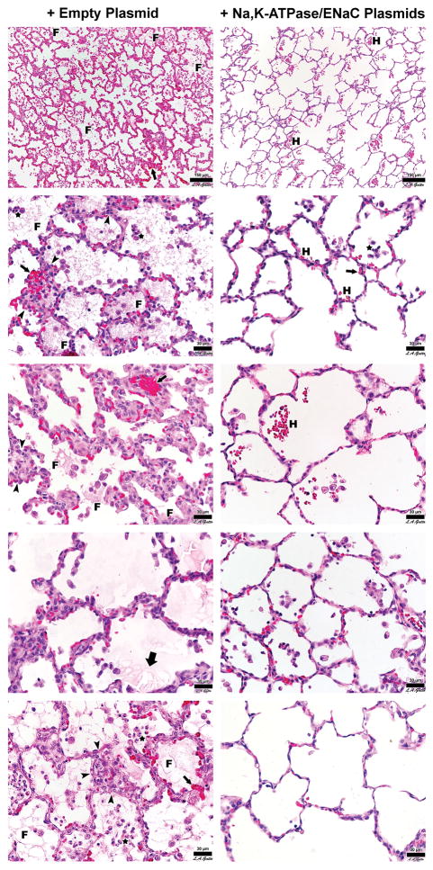
Figure 7. Representative H&E stained histology sections of lung tissue.
Representative photomicrographs of lungs from treatment plasmid or empty plasmid groups are shown at high magnification. Each panel represents a different animal. Sections of the lungs were chosen at random for each animal and are representative of the degree of injury found throughout the lungs. F = fibrin deposit, H = hemorrhage, Small Arrow = alveolar wall thickening, Thick Arrow = alveolar edema, Arrowhead = leukocyte infiltrates, Star = intra-alveolar cell infiltrates.

