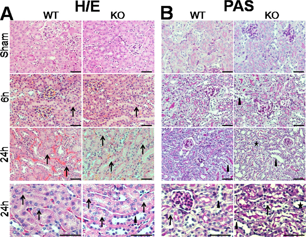
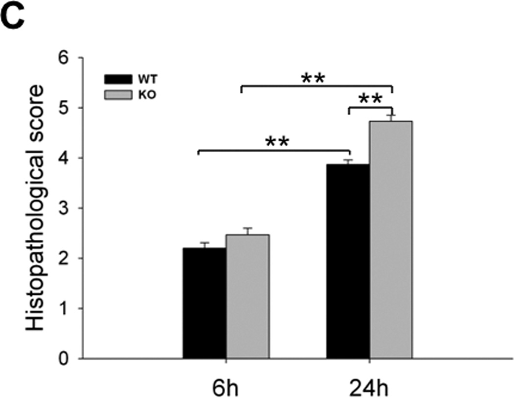
Fig. 2. Histology of the kidney in septic WT and SP-A/SP-D KO mice.
The histopathology of kidney tissues was analyzed in septic WT and SP-A/SP-D KO mice, as well as control (sham). Histological sections of kidney tissues were stained with H/E (A) or PAS (B) method. The histological score of kidney tissue injury was assessed (C). Kidney histology shows the presence of vacuolar degeneration of tubular cells (arrows), flattened tubular cells with tubular lumen dilatation (stars) and lack of brush border (triangles) in septic but not sham mice. Compared with septic WT mice, the kidney injury score is higher in septic SP-A/SP-D KO mice 24 h after CLP. Magnification ×200 or ×400. Graphs represent the mean ± SEM. **p<0.01. Bars = 100 µm. (n = 6 mice/group)


