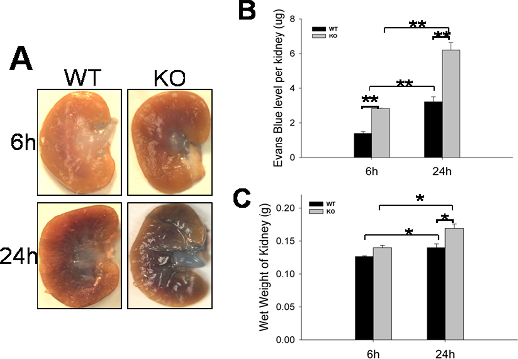
Fig. 3. Kidney vascular permeability and wet weight of the kidneys in septic WT and SP-A/SP-D KO mice.
Evans Blue (EB) dye (50 mg/kg) was injected via the mouse tail vein and EB leakage as described in the methods. Representative photos of the kidneys from septic WT and SP-A/SP-D KO mice 6 h or 24 h after CLP are shown in the panel (A). The levels of EB dye (B) recovered in kidney tissues and the wet weights of the kidneys (C) were higher in septic SP-A/SP-D KO mice compared with septic WT mice. Values represent the mean ± SEM, *p<0.05, **p<0.01. (n = 6 mice/group)

