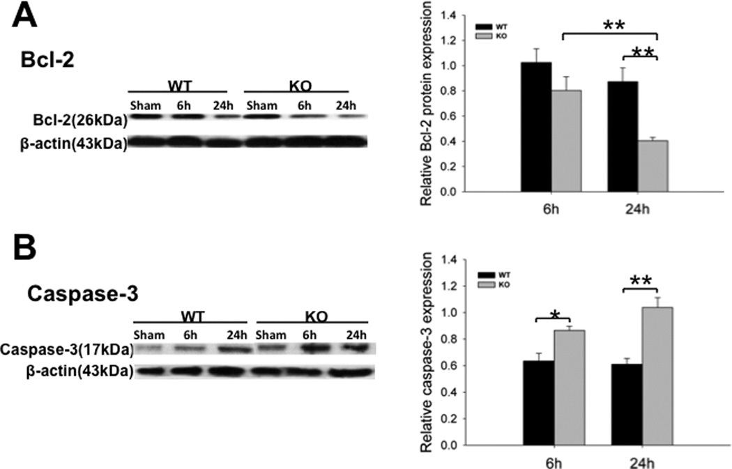
Fig. 5. Expression of Bcl-2 and caspase-3 in the kidney of septic WT and SP-A/SP-D KO mice.
Protein levels of Bcl-2 (A) and activated caspase-3 (B) in the kidney were determined by Western blotting analysis and quantified by densitometry. Protein levels were normalized to β-actin. Decreased level of Bcl-2 and increased level of activated caspase-3 were found in septic SP-A/SP-D KO mice compared to septic WT mice 6 h and 24 h post-CLP. Graphs represent the mean ± SEM, *p<0.05, **p<0.01. (n = 6 mice/group)

