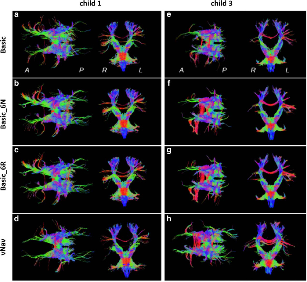
Figure 7.
Tractography reconstructions for fibers passing through the brainstem for two children. Axial projections (anterior is on the left) and coronal projections (anatomic left is on the right) are viewed from superior and anterior locations, respectively, for various reconstructions: (a,e) Basic scan, no retrospective correction; (b,f) Basic_6N; (c,g) Basic_6R; and (d,h) vNav. Coloration is by local spatial orientation: left–right (red); anterior–posterior (green); superior–inferior (blue).

