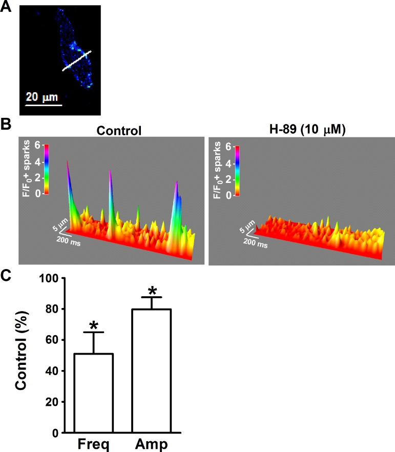
Fig. 3.
Pharmacological inhibition of PKA with H-89 suppressed the basal level of Ca2+ sparks in freshly isolated UBSM cells. A: an image of a freshly isolated UBSM cell loaded with fluo 4-AM. The white line passing through the active site a is the laser beam-scanning pathway (1-pixel width). B: the 3-dimensional view of the recordings illustrate the Ca2+ sparks in the absence and presence of H-89 (10 μM). The color scale indicates the relative fluorescence intensity F/F0. C: summary data illustrating that H-89 (10 μM) decreased the Ca2+ spark frequency (Freq) and amplitude (Amp) in UBSM cells (n = 8, N = 6) *P < 0.05.

